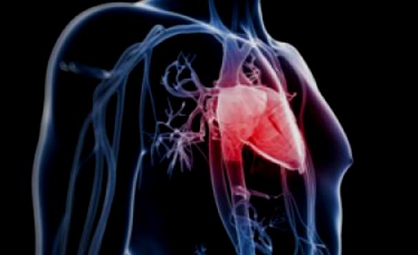
So far, it has been impossible to reliably estimate the risk of a patient to suffer from atrial flutter. Researchers of Karlsruhe Institute of Technology, the Medical Clinic IV of the Karlsruhe Municipal Hospital, the Faculty of Medicine of the University of Freiburg, and the University Heart Center of Freiburg-Bad Krozingen have now developed a method to assess the individual risk of atrial flutter. As reported by the scientists in Frontiers in Physiology, personalized computer models enable identification of all pathways along which atypical circulating electric excitations may occur.
Our models consider anatomic, electrophysiological, and pharmacological criteria."
Dr. Axel Loewe, Head of the Computational Cardiac Modeling Group of KIT's Institute of Biomedical Engineering (IBT)
In this way, the effect of therapies, such as catheter ablation or medication, can also be assessed in advance.
The work reveals the advantages of mathematically simulated organs for medicine: "Computer models offer a perfectly controllable environment for experiments," Loewe says. "Single modifications can be modeled and their effects on the complete system can be calculated." These models supplement classical methods, such as cell and animal experiments, and enable tests of new therapies without any risks for humans.
In his doctoral thesis, Loewe already modeled the causes of atrial flutter on the computer. The Computational Cardiac Modeling Group headed by Loewe develops close-to-reality models of the heart on all levels, from the ion channel to cells and tissue, to the complete organ. In this way, they can model how an electric excitation develops, spreads to the cardiac atria and the entire heart, and stops in a healthy heart or is self-sustaining in case of cardiac arrhythmias.
Apart from modeling such fundamental physiological and pathological processes, the group also develops personalized models to individually determine the risk of diseases and the effect of treatments. To capture the patient's individual anatomy, such as the size and shape of the atria, the researchers use imaging methods, such as magnetic resonance imaging. To integrate the ECG-recorded electrical activity of the heart, the group closely cooperates with IBT's Bioelectric Signals Group that is headed by Professor Olaf Dössel. Work at the interface of engineering, computer science, natural sciences, and medicine paves the way towards customized therapies.
Atrial flutter is an abnormal heart rhythm, in which unusually fast electric excitation patterns cause the cardiac atria to contract quickly. Contrary to the more common atrial fibrillation, electric excitation in case of atrial flutter is coordinated. But as atrial fibrillation, atrial flutter may cause the heart to beat too fast, breathing difficulty, and weakness. Moreover, the risk of a stroke is increased.
A typical treatment of atrial fibrillation is ablation, i.e. catheter-supported obliteration of pathogenic electric excitation sources in the myocardial tissue. After the treatment, however, patients frequently develop so-called atypical atrial flutter, in the case of which a circulating excitation may occur both in the left and in the right atrium.
Karlsruhe Institute of Technology
Journal reference:
Loewe, A. et al. (2019) Patient-Specific Identification of Atrial Flutter Vulnerability–A Computational Approach to Reveal Latent Reentry Pathways. Frontiers in Physiology. doi.org/10.3389/fphys.2018.01910.


No comments
Post a Comment