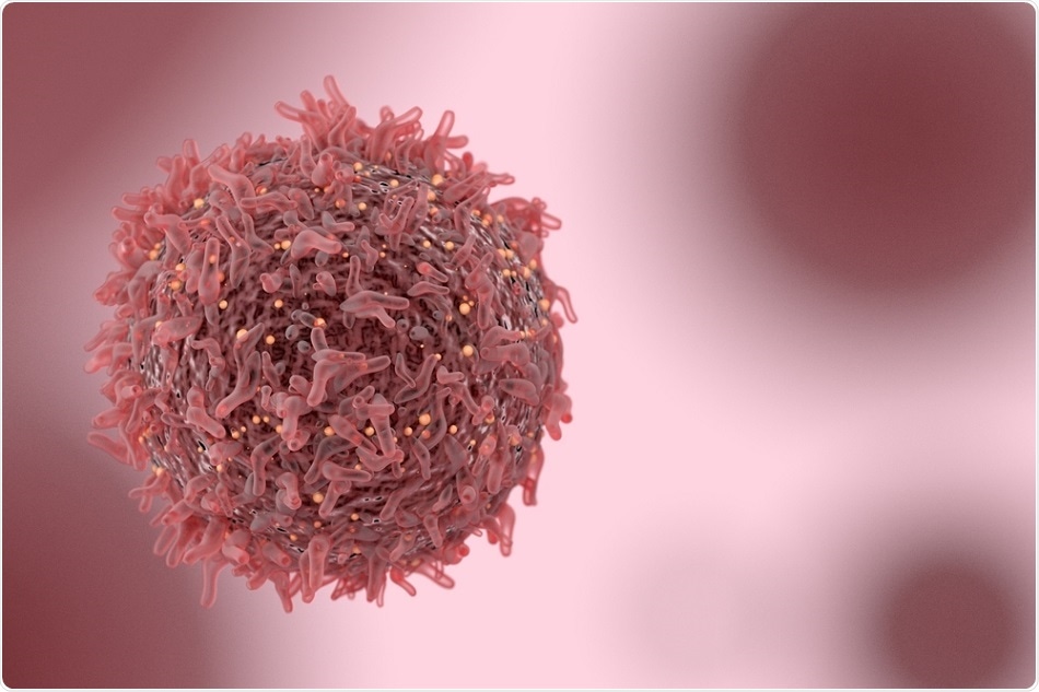
Groundbreaking study uncovers activity of senescent cells
Researchers at the Tulane School of Medicine, New Orleans, LA, have recently published a paper revealing their findings from a study they conducted, which looked into the activity of senescent cells. Their data uncovered that senescent cells induced by chemotherapy can remain persistent through engulfing the surrounding cells.They observed that in chemotherapy-treated breast cancer, wild-type p53, a tumor suppressor protein, coerces cells into senescence, rather than apoptosis. This means that cells enter a state where they no longer divide or grow, but they do not enter the usual programmed form of cell death. This mechanism induces relapse through the senescence-associated secretory phenotype.
The team, led by Crystal Tonnessen-Murray, watched how the cells interacted within treated tumors via time-lapse and confocal microscopy. They noticed that neighboring non-senescent tumor cells or senescent cells were enveloped by chemotherapy-induced senescent cells.
Following this, they observed the enveloped cells processed through the lysosome and subsequently broken down. The process enabled the chemotherapy-induced senescent cells to persist.
Further to this, the team ran a gene expression analysis, which uncovered that chemotherapy-induced senescent cell lines and tumors demonstrated an increased up-regulation of conserved macrophage-like program of engulfment. These findings show how senescent cells can endure, and how they can influence relapse in tumors that respond worst to therapy.
What are senescent cells?
Previously, it has been accepted that cells that enter senescence can prevent cancer growth through its prevention of cell division. Now, scientists have been able to show that in some scenarios, senescent cells can act as a catalyst in cancer progression via secretion of growth factors and immune-signaling molecules.
Chemotherapy and senescence
Senescence is triggered by chemotherapy due to the damage the treatment causes to the DNA of the cells. This damage ideally leads to cell death, but senescence is another possible outcome.The work from the LA-based team showed that cells that become senescent have the capability of causing more damage. They observed senescent cells ingest neighboring cells, and process them through the lysosome, the cell’s digestive organelle. Following this, they found that these cells that engage in cannibalism had increased longevity compared with those that had not ingested cells, meaning that this destructive activity promotes their survival. The researchers suggest that this enhanced survival was a result of obtaining metabolic building blocks from the lysosomal digestion.
While this is not the first time that cell cannibalism has been reported in cancer, this was the first study that was able to link it with senescence. For the first time, evidence was able to show that the link between cannibalism and senescence may be fundamental to the endurance of senescent cells within cancer tissues.
Future research directions
The result of this study was surprising as cell death has not previously been seen to enhance the survival of senescent cells. The study implicates the importance of understanding the complexity of cell death regulation in multicellular animals, particularly in cancer. To go ahead and create new and more effective treatments for cancer, it is essential that further research is carried out in this field in order to uncover more about the properties that support cancer cell survival and relapse following chemotherapy.Journal reference:
Chemotherapy-induced senescent cancer cells engulf other cells to enhance their survival. Journal of Cell Biology. DOI: 10.1083/jcb.201904051. Published on September 17, 2019. http://jcb.rupress.org/content/early/2019/09/16/jcb.201904051


No comments
Post a Comment