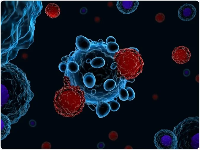
A new Finnish study suggests that type 1 diabetes may in part be caused by a novel subset of immune cells. This is because these cells, called peripheral T helper cells, are found to circulate at a higher rate in children with type 1 diabetes of recent onset, as well as in healthy children who later develop the condition. Both T and B cells are immune cells belonging to the category called lymphocytes, which are crucial for cellular and antibody-mediated attack of foreign cells and particles.

Type 1 diabetes is an autoimmune disease that causes loss of blood sugar control. It is caused by the immune destruction of the insulin-producing clusters of beta cells in the pancreas, called the islets of Langerhans. It typically shows up first in childhood, but doesn’t produce any symptoms at first. However, even at this stage it is detectable by finding autoantibodies to the islet cells. When two or more islet cell autoantibodies are present the individual has at least a 50% risk of developing type 1 diabetes within five years.
We already know that the B cells responsible for generating these antibodies against proteins in the islets are being driven by T cells called follicular T helper cells (Tfh). Previous research by the same scientists has shown that the number of Tfh cells in blood is increased in type 1 diabetes, soon before the disease becomes evident. Moreover, this is seen only when more than one type of autoantibody (AAb) is present.
The current study focused on the role played by a newly described type of T cell called C-X-C motif chemokine receptor type 5-negative, programmed cell death protein 1-positive (CXCR5−PD-1hi) peripheral T helper (Tph) cells.
To do this, the researchers used a method called color flow cytometry to find out whether these Tph cells were responsible for promoting B cell antibody production like Tfh cells. They also measured the number of Tph cells in circulation in three sets of children:
- children with recently diagnosed type 1 diabetes
- children who were positive for autoantibodies (AAb+) but not yet diabetic
- children who neither had AAb nor showed evidence of disease
What were the results?
The analysis showed that these cells behaved much like Tfh cells, but showed an important difference: instead of having receptors that home in to lymph nodes like Tfh cells, they have chemokine receptors that attach them to areas of inflammation. This could mean that they drive the B cell’s antibody production in the inflamed pancreas.
Secondly, the numbers of both Tfh and Tph cells were higher in the first set of children, but only if they had more than one autoantibody. Finally, Tph number was also increased in some of the second group of AAb+ disease-negative children. On follow-up, the researchers found that the children with higher Tph cell frequency in circulation were those who subsequently developed type 1 diabetes. The findings were validated in another comparison between children with newly diagnosed type 1 diabetes and children who did not.
The current study also found a molecule called TIGIT that is present on the cells of the Tph lineage to be useful in identifying the exact type of these cells that is linked to the occurrence of autoimmunity. More questions remain as to whether the Tph and Tfh cells are themselves responsible for the damage to the beta cells, or are just produced by bystander T cells not involved in the autoimmunity.
The number of Tph cells in blood is very low, and so is the magnitude of the increase in these cells in circulation. However, it is quite possible from the earlier study on Tfh cells that these cells are present in much greater abundance in the inflamed islets in the pancreas. Advanced studies are required to test the concept that Tph cells may partly cause the pancreatic beta cell destruction.
The researchers conclude that an increase in the number of Tph cells in circulation is linked to a higher risk of developing type 1 diabetes. This confirms earlier studies showing that T and B cells are vitally linked in the development of this condition.
Researcher Ilse Ekman says, “Based on our results, it is possible that peripheral helper T cells may have a role in the development of type 1 diabetes. This information could be employed in the development of better methods to predict type 1 diabetes risk and new immunotherapies for the disease.”
The study was published in the journal Diabetologia on July 3, 2019.
Ekman, I., Ihantola, EL., Viisanen, T. et al. Circulating CXCR5−PD-1hi peripheral T helper cells are associated with progression to type 1 diabetes, Diabetologia (2019) 62: 1681. https://doi.org/10.1007/s00125-019-4936-8, https://link.springer.com/article/10.1007%2Fs00125-019-4936-8


No comments
Post a Comment